Rickets in infants
At present, ecological problems and wrong nutrition frequently provoke hypovitaminosis D in infants. Due to complexity and limited practical availability of direct methods for early diagnosis of hypovitaminosis D in healthcare institutions indirect diagnostics are adopted which are not so plausible. This results in a late discovery of rickets in infants.
Various perinatal factors (mother's disease during pregnancy, unfavorable course of labor etc.) also create preconditions for rickets.
Due to the fact that intensive transfer of calcium and phosphorus from mother to fetus occurs during the last months of pregnancy, premature infants already at birth frequently have osteopenia. This medical term means low content of mineral substances in bones. Irrational nutrition and irregular life regimen of the pregnant woman aggravate the situation. Development of rickets in infants is provoked by the immaturity of enzymatic systems of liver, kidneys, skin as well as the related diseases. Rickets especially often affects premature children. For a normal ossification process, food should contain sufficient amounts of proteins, calcium and phosphorus combined in a correct ratio, trace elements such as magnesium and zinc as well as vitamins B and A.
Lack of physical activity of the child leads to rickets, since supply of bones with blood and electrostatic tension significantly improve when muscles are active.
Signs of rickets
At the initial stage (during the first year of infant's life) changes of the nervous and muscular system are stated.
The child gets irritable, in certain cases anxious, he starts at loud sounds and bright light and he sleeps uneasily. Hyperhidrosis, especially on the head, and baldness on the neck occur. 2 to 3 weeks later morbidity of bone edges in the region of the big fontanel, along arrow-shaped and lambdoid sutures is detected. Muscular tonus relaxes. Calcium content in blood remains normal, while phosphorus content drops insignificantly. Urine tests show phosphaturia.
When the disease reaches its highest point, symptoms related to the nervous and muscular system prevail. Sweating, weakness, muscle and ligament hypotonia increase, lagging in psychomotor development is noticeable. This period is characterized by rapidly progressing bone changes: malacia of plane cranial bones, craniotabes, hind head flattening, head asymmetry. Excrescence of osteoid tissue in points of plane cranial bone ossification results in formation of frontal and occipital tubers. Due to this the head obtains a square or gluteal form. Deformations of facial regions of the skull can also occur, such as saddle nose, “olympic” front, occlusion abnormalities etc. Teeth erupt with a delay and non-consecutively, they are easily affected by caries.
The chest is often deformed. On ribs, in union points of union of cartilage and osteal parts tubercles appear. Chicken breast, rickets kyphosis, lordosis and scoliosis can potentially develop. A deep retraction ( Harrison 's groove) is formed outside the chest on the level of diaphragm attachment. Rib edges of inferior aperture are turned forward like brim of a hat due to big stomach size.
Prime clinical signs of rickets are bone changes.
Head :
craniotabes occurs in the occipital or parietal region. There, the skull is so soft that it can be compressed.
delayed eruption of teeth
Chest:
Rickets tubercles develop as a result of cartilage hypertrophy between ribs and the breast bone which have the form of an intumescence located on both sides of the breast bone.
chest deformation :
Spine:
Spine bone changes occur in absence of normal bowing and of pathological curvatures in form of kyphosis, lordosis and scoliosis
Extremities :
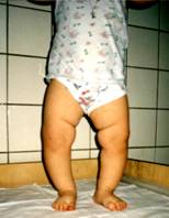
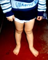
Abnormalities in hip joint and inferior limb development occur toward the end of the first and the beginning of the second year of baby's life in form of: bandy legs (see figure), baker's legs (see figure), flat rickets pelvis.
A concomitant sign is muscular weakness. Frequent respiratory infections are also characteristic, as well as asiderotic anemia of various severity and latent anemia. Children having rickets are often affected by changes in other organs and systems besides the bone system. Muffled heart sounds, tachycardia, systolic murmur, development of atelectatic regions in the lugs and protracted pneumonia, large liver and spleen mass are to be listed among further negative phenomena. Development of conditioned reflexes delays, while acquired reflexes weaken or entirely disappear.
Treatment
Treatment of infants affected by rickets should be aimed at the elimination of vitamin D deficiency, normalization of phosphoric and calcium metabolism, elimination of acidosis, activation of osteogenesis and nonspecific correction.
Drug therapy of rickets in infants consists in administration of vitamin D. Two types of vitamin D are prescribed for children: vitamin D2 (ergocalciferol) of herbal origin and vitamin D3 (cholecalciferol) of animal origin. Their chemical composition differs . Cholecalciferol is considered to be the prevailing drug.
Cholecalciferol is prepared in form of aqueous solution “AQUA – D3” and oil solution “Vigantol” for intake.
Vitamin D3 (other names: Cholecalciferol, Aqua D3, Vigantol)) is to be taken once a day 10 drops for 45 days for infants aged under one year. Older children should take 10 drops of vitamin D3 once a day for 45 days. Three therapy sessions with intervals of 2 months are required (see in “Medicaments” by M.Mashkovsky, 2005, pp.636-639).
Combined signs of hypervitaminosis are lack of appetite, vomiting, frequent urination. Causes of disturbance disappear immediately after discontinuation of taking vitamin D.
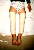
Should symptoms of bone rickets be discovered in an infant he must be subject to a checkup by an orthopedist. Bone deformations in children aged under one year can be corrected relatively easy without adoption of special treatment methods, only by taking vitamin D 3 . At an older age deformations of bones, in particular those in lower limbs, increase dramatically so that an urgent treatment is necessary. In children aged under 4 deformations of lower limbs can be corrected through adoption of conservative treatment methods for 2 to 3 months. Correcting plaster bandages are used for this purpose. They are applied on the infant's legs under fixation of knee joint. The foot remains free so that the infant can walk. Children get accustomed to the bandage very soon (it takes a day or two as a rule), after that the bandage does not cause any inconvenience with them.
Older children need to be operated on in order to eliminate a deformation.
Dear parents ! Take good care of your children . Do not neglect their diseases.
Up
|