Platypodia
It is one of the most frequent foot deformations in infants. The term “platypodia” denotes deformity of all foot's arches. Flattening of the longitudinal foot arch is known as longitudinal platypodia, while flattening of the transverse arch is called transverse platypodia. Main support elements of the foot's arches are front and rear tibial muscles of the lower leg as well as short and long flexor muscles of toes. If those muscles become weak, the child's foot flattens under the body weight and turns outwards at the start of standing or walking. The inner edge of the foot rests on the floor, the forefoot is abducted outwards.
Actual platypodia can be diagnosed only after the completion of infant's growth. Prior to that period the child's foot is in the process of formation. This condition is characterized by orthopedists as planovalgus deformity or foot setting.
Preventive treatment started at the initial stage of such deformity guarantees with a good probability healthy foot formation in the infant.
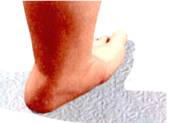
Planovalgus deformity of the foot can be discovered in the first months of the child's life. To do this, press the child's foot outwards with your thumb to the maximum extent possible. In case of planovalgus deformity, the child's foot leans with its back against the front surface of the leg and is turned outwards shaping a prominence on the inner edge.
Affected children, when they start walking, draw in toes and shuffle with one foot in walking.
In schoolchildren, platypodia causes pain and fatigability in muscles of the lower leg during walking and toward the end of the day. Further on, pain increases at standing and walking. School results become poor, headache and quick general fatigability appear. Pain is usually localized on the sole, in the arch region and in lower leg's muscles.
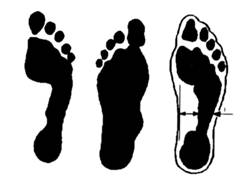
Platypodia can be diagnosed by plantography (footprint) method. To do this, apply some vegetable oil on the foot's sole and put the foot on a clean sheet of paper. You will get a footprint on which you can determine the condition of the foot's arch.
With regular form, the neck on the inner edge of the foot is equal to 2/3 of its width. In case of platypodia, such a neck is very small or not present at all.
The treatment of platypodia in an infant should commence in the first months of his life. At this age it is necessary to eliminate muscle weakness and laxity of foot's and lower leg's ligaments. Massage, remedial exercises and physiotherapeutic procedures can be adopted as treatment methods.
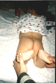
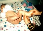
Massage of lower leg's and foot muscles can be done by parents themselves. Sometimes, a work-up of the child's leg muscles with fingers would suffice. To massage foot's muscles, pass your thumb over the foot's sole from toes to the heel. Massage should be performed twice a day (in the morning and in the evening) for 10 minutes.
At the age of 1 year, when the child starts to walk, remedial exercises should be intensified. You should learn him walk and jump on his toes, walk on the outer edge of the sole, on heels, roll a pencil or a round stick with the foot and use a massage roller.
To strengthen lower leg's and foot's muscles, 10 sessions of electrical myostimulation of front and rear tibial muscles and of the short and long flexor muscles of toes are adopted. 4 courses of electrical myostimulation are needed annualy.
In preschool children, supinators and orthopedic shoes should not be used in case of static platypodia. Fixation of the foot can provoke weakening of leg's and foot's muscles and worsen the foot's condition.
Treatment of platypodia in schoolchildren should be performed as follows:
Treatment of planovalgus deformity and platypodia
- Therapeutic exercises on a Stepper training device for 20 to 30 minutes a day.
- Massage of lower leg's and foot's muscles: 10 sessions x 4 times a year.
- Physiotherapy: 4 courses a year.
- Electrical stimulation of the long and short flexor muscles on both feet: 10 sessions per foot.
- Electrical stimulation of the front and rear tibial muscles: 10 sessions per leg.
Checkup by a pediatric orthopedist every 6 months.
In case of pain and fatigability in infant's legs at walking and standing supinators should be used. They maintain the foot's arch in the correct position and prevent pain in feet.
Up
|